Structure of Phage Virus with Diagram
Earlier studies were made on the phage virus (Bacteriophage). This virus infects Escherichia coli (bacteria). There are different types of phage. Mostly T phages (T for type) were studied T2 and T4 are mainly used in phage studies. The structure of T4 is studied by an electron microscope.
The volume of phage is about 1\1000 of the host (bacteria). It resembles tadpole (frog larva). It consists of head and tail).
Head of Phage Virus: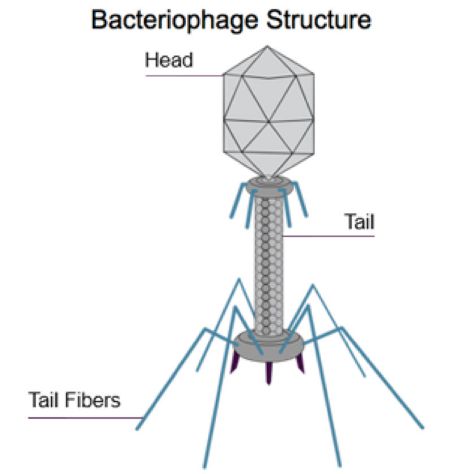
It has elongated head. Head has different shapes like, pyramidal, hexagonal and prism-shaped structure. Tail is attached with the head. Double stranded DNA molecule is present inside the head.
Tail of Phage Virus:
Tail has complex structure. A layer of protein forms the inner tube or core of the tail. This core is enclosed in a sheath. This sheath is made up of another type of protein. A collar is present on one side of sheath.
The other side of sheath has end plate. Six tail fibers are attached with the end plate. Tail fibers are structures of attachment.
Replication of Bacteriophage:
The Bacteriophage or phage virus replicates only inside the bacterial cell. Phage virus shows two types of cycles during its replication:
[wp_ad_camp_2]
1. Lytic cycle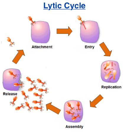
The phage virus causes the lysis of the bacteria in the lytic cycle. Such viruses are called lytic or virulent phage. Lytic cycle is divided into following steps:
i. Attachment or adsorption
The Bacteriophage is attached at the receptor site on the cell wall of the bacterium. Weak chemical union takes place between the virus and the receptor site.
ii. Penetration
The tail of virus releases lysozyme enzyme. It dissolves a small part of the bacterial cell wall. The tail sheath contracts and pushes the tail core into bacteria through its cell wall and cell membrane.
The virus injects its DNA in to the bacterial cell like syringe. The protein coat (tail and head) of the virus remains outside the bacterial cell. However, many animal viruses enter the host as a whole.
[wp_ad_camp_3]
iii. Multiplication
Inside the bacterial cell, the viral DNA starts controlling the biosynthetic machinery of the bacteria. It induces the bacterial cell to synthesize the parts of the virus, protein and DNA. Then virus multiplies to form many new viruses. Approximately 200 new viruses are formed after 25 minutes of the infection.
iv. Lysis
Many viruses are formed in the bacteria. The bacterial cell undergoes lysis and bursts. The newly formed viruses are released and infect other bacteria.
2. Lysogenic Cycle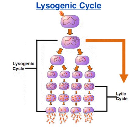
The phage does not always destroy bacteria. Sometimes it lives peacefully, inside the bacterial cell. It does not control the biosynthetic machinery of the host bacteria. The viral DNA incorporated into the bacterial chromosome.
The phage in this state is called Proghage. This process is called lysogeny. The bacteria continue to live and reproduce normally in this condition. The viral DNA becomes the part of the bacterial chromosome. It passes to each daughter cell in all coming generations.
Sometimes, the viral DNA detaches from the bacterial hromosome and starts lytic cycle. This process is called induction. The sogenic bacteria are resistant to viral infection. The phage which causes sogeny is called temperate or lysogenic phage.